01
New chest discomfort, breathlessness, palpitations or fainting
↗
Ultrasound assessment of heart structure, valves and function.
Worried about cost? You'll get a written estimate before any planned procedure.
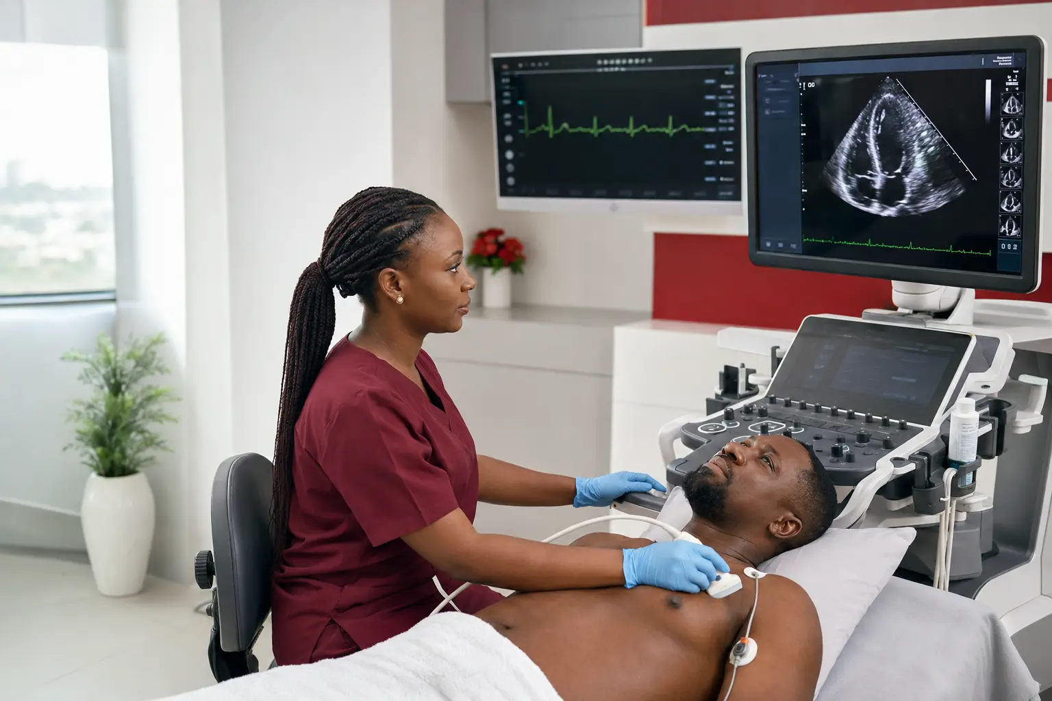
An echocardiogram provides moving images of the heart and helps clinicians assess chambers, pumping function, valves, fluid and other important findings.
Cardiovascular tests can show heart structure, rhythm, circulation, blood-pressure patterns and response to activity. We start with the clinical question, select an appropriate investigation and explain what the result does—and does not—mean for the next decision.
Testing should reduce uncertainty, not create more confusion. We explain why the test was requested, what to expect and how the result connects to your care plan.
These are common starting points, not a diagnostic checklist. The right assessment depends on the full clinical picture.
New chest discomfort, breathlessness, palpitations or fainting
↗An abnormal examination, ECG or laboratory result
↗Monitoring a known heart or valve condition
↗Planning a procedure or operation
↗Checking response to treatment
↗Clarifying conflicting or incomplete previous results
↗Every capability is selected according to clinical need. Your team should explain why a test, treatment or referral is being considered.
A transthoracic echocardiogram images heart chambers, valves and pumping function in adults.
→Paediatric echocardiography assesses heart structure and function in children, including congenital findings.
→Doppler ultrasound measures blood-flow speed and direction across the heart's valves and chambers.
→Echocardiography assesses heart function before surgery or a major procedure to guide anaesthetic and surgical planning.
→Images and measurements are interpreted by a cardiologist rather than left as raw data.
→Complex medicine feels more manageable when every stage is visible and every question has a place.
A clinician identifies what needs to be confirmed, excluded or monitored.
The team explains clothing, fasting, medicines or activity instructions where relevant.
Trained staff obtain the measurements and images needed for reliable interpretation.
The result is read in clinical context rather than treated as an isolated number or image.
The report feeds into follow-up, treatment, another test or reassurance when no further action is needed.
An echocardiogram uses high-frequency sound waves to create moving pictures of the heart. A routine transthoracic echo is non-invasive and does not use ionising radiation. It can show chamber size, pumping function, wall movement, valve performance, blood-flow patterns and fluid around the heart.
It helps assess murmurs, enlarged heart, unexplained breathlessness or chest symptoms, hypertension-related changes, valve disease, heart failure and congenital conditions.
You usually lie on an examination couch while gel and a handheld transducer are placed on the chest. Sticky ECG electrodes may be added to record rhythm during the study.
A straightforward study may be completed quickly, while complex valve, congenital or heart-function questions require more views and measurements.
Selected patients may need stress echocardiography or transoesophageal echocardiography to answer a question that a routine chest study cannot show clearly.
These are common reasons to arrange an assessment. They do not confirm a diagnosis, and the appropriate starting team may change after clinical review.
Cardiocare brings the relevant specialists, investigations, treatment and follow-up into one documented plan.
The referral and clinical history tell the sonographer and cardiologist which structures and measurements need particular attention.
→Height and weight may be recorded and clothing is adjusted privately so the transducer can reach the left side of the chest.
→Images and Doppler measurements are taken from several positions. The test is usually painless, although gentle pressure may be needed for a clear view.
→A cardiologist reviews structure, pumping function, valves and flow patterns in the context of the clinical question.
→Findings can move directly into cardiology follow-up, intervention, cardiac surgery, heart-failure care or reassurance.
The strongest advantage is not one department. It is the ability to bring the right departments together quickly and safely.
Results are interpreted within a hospital that can provide cardiology consultation, rhythm testing, coronary assessment and long-term follow-up.
Echo findings help cardiac surgeons, interventional cardiologists and anaesthesia teams prepare for selected procedures and operations.
Cardiocare supports adult studies and connects paediatric questions to the appropriate specialist pathway within The Limi Hospital Group.
Results can be reviewed within an immediate cardiology, intervention or surgical pathway where needed. This is why patients, families and referring clinicians choose Cardiocare for care that may need more than one specialist or facility.
Consultation, diagnostics, intervention, surgery, ICU/HDU, dialysis and recovery can coordinate without fragmenting the patient journey.
Advanced catheter, device and surgical expertise—including Northern Nigeria’s first cardiac left bundle branch pacing—reflects a culture of clinical progress.
Round-the-clock emergency, ICU and HDU support surrounds patients whose needs may change quickly.
Cardiology works alongside nephrology, endocrinology, neurology, pulmonology, surgery and other specialists when one diagnosis is not the whole story.
International-level cardiovascular capability in Abuja helps families avoid unnecessary travel abroad and remain closer to their support system.
Plain-language teaching, patient guides, support programmes and professional education turn expertise into confidence.
Open each question for a straightforward explanation. Your own clinician should still interpret what it means for you.
Ask the Cardiocare teamNo. Some findings are minor, temporary or need comparison with symptoms and other tests before their importance is clear.
Sometimes, but many cardiovascular questions need several pieces of information—history, examination, ECG, imaging or laboratory results.
The clinical team should review the finding with you and explain the next step. Timing depends on the test and urgency.
If a result needs specialist review, intervention, surgery or monitored care, those pathways are already connected.
Prepare questions, understand warning signs and revisit explanations at your own pace.
This page is educational and cannot diagnose a condition or determine which treatment is right for an individual. A clinician must assess symptoms, results, medicines and overall health. Severe or rapidly worsening symptoms need urgent care.
Emergency? Go to the nearest emergency department — do not drive yourself. Our cath lab is on standby 24/7.
Call 24/7: +234 806 142 4614