
Echocardiography: what an ultrasound of the heart can show
An echocardiogram uses sound waves to examine heart structure, valves, blood flow and pumping function.
Understanding echocardiography
An echocardiogram (often shortened to "echo") is an ultrasound scan of the heart. A handheld device called a transducer sends painless, high-frequency sound waves into the chest; these waves bounce back differently depending on what they meet — heart muscle, blood, or a valve — and a computer turns the pattern of echoes into a moving picture of the beating heart in real time. Unlike an X-ray or CT scan, echocardiography uses no ionising radiation at all, which is one reason it can be repeated as often as needed to follow a condition over time.
An echo can show the size and shape of each heart chamber, the thickness and strength of the heart muscle, how well each of the four valves opens and closes, and — using a technique called Doppler — the speed and direction of blood flow through the heart. Together, this makes echocardiography one of the most informative single tests in cardiology: it can help explain a murmur, breathlessness, ankle swelling, palpitations, high blood pressure affecting the heart, suspected heart failure, valve disease or an enlarged heart. Selected questions that a resting scan cannot fully answer may call for stress echocardiography, which adds exercise or medicine, or a transoesophageal echocardiogram, which images the heart from inside the gullet for a closer view.
When an echocardiogram is recommended
Your clinician may recommend an echo for many reasons, including:
- ●A heart murmur or other abnormal finding on examination
- ●Breathlessness, ankle swelling or reduced exercise capacity
- ●Known or suspected valve disease or heart-muscle disease (cardiomyopathy)
- ●Longstanding high blood pressure, to check whether it is affecting the heart
- ●Palpitations, to look for a structural cause
- ●Monitoring heart function before or after a procedure, surgery, or certain cancer treatments
- ●A family history of heart-muscle disease, as part of screening
How to prepare for your scan
Preparation is usually straightforward, but the details differ slightly by scan type:
- ●For a standard (transthoracic) echo, no fasting is needed and you can take your usual medicines — eat and drink normally beforehand
- ●Wear a two-piece outfit or clothing that allows easy access to your chest
- ●If a transoesophageal echo (TOE) has been arranged, you will usually be asked to fast for several hours beforehand, and you must arrange for someone to accompany you home, as sedation is normally used
- ●If a stress echo has been arranged, wear comfortable clothing and footwear suitable for light exercise, and ask whether any regular medicines should be paused beforehand
- ●Bring your referral letter and any previous echo or heart reports for comparison
What happens during the scan
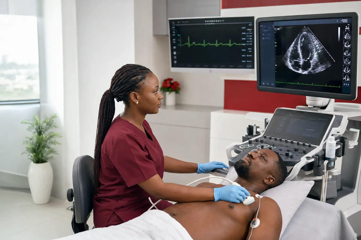
For a standard transthoracic echo, you lie on a couch, usually turned slightly onto your left side, with your chest exposed. A clear gel is applied to the skin to help sound waves travel between the transducer and the body, and the sonographer moves the transducer gently over several positions on the chest to capture different views of the heart. You may be asked to hold your breath briefly, breathe in a particular way, or shift position during the scan. The room is often kept dim to make the ultrasound images easier to see on the screen. The scan itself is painless, though the gel can feel cool, and it typically takes some time to capture all the required views thoroughly.
Stress echocardiography
For a stress echo, images are taken at rest and then again immediately after your heart rate has been raised — either by exercising on a treadmill or stationary bicycle, or with a medicine that has a similar effect if you are unable to exercise. Your heart rate, blood pressure and ECG are monitored throughout, alongside the images, to see how the heart responds under demand.
Transoesophageal echocardiography (TOE)
For a TOE, your throat is numbed with a local anaesthetic spray and you are given sedation to help you relax. A thin, flexible probe is then gently guided down the gullet, which lies directly behind the heart, giving a closer and often clearer view of certain structures — particularly the heart valves and the upper chambers — than a scan from outside the chest can provide.
How the scan feels
A standard transthoracic echo is painless; you may feel firm but gentle pressure from the transducer, and the room may feel cool. A stress echo will make you feel your heart working harder, similarly to normal exercise or exertion, and you may become breathless — this is expected and monitored throughout. A TOE involves some throat discomfort as the probe is swallowed, similar to the sensation of a dental impression, though the sedation and throat spray make this manageable for most people; many people remember little of the procedure afterward because of the sedation.
Getting your results
The images and measurements are reviewed and reported by a cardiologist. Your referring clinician will usually discuss the findings with you at a follow-up appointment, where the result can be explained in the context of your symptoms and overall health — ask your care team when and how you can expect to receive your result if this is not clear at the time of your scan.
Risks to know about
Echocardiography is one of the safest tests in cardiology:
- ●A standard transthoracic echo carries no known risk — it uses only sound waves, with no radiation or injection involved
- ●A stress echo carries the same small risks as any supervised exercise or stress test, and is stopped immediately if concerning symptoms appear; it is performed with monitoring throughout
- ●A TOE carries a small risk related to the sedation and the passage of the probe, including throat irritation or, uncommonly, minor injury — your team will discuss this with you specifically if a TOE is recommended
Recovery and aftercare
Recovery needs depend on which type of echo you have had:
- ✓After a standard transthoracic echo, you can resume normal activities immediately, including driving
- ✓After a stress echo, you will be monitored briefly until your heart rate and blood pressure return to normal before you leave
- ✓After a TOE, you will be observed until the sedation wears off, you should not drive, operate machinery or make important decisions for the rest of that day, and you may have a mildly sore throat for a short time afterward
- ✓Keep your follow-up appointment to discuss the result, even if you feel entirely well
Do not wait if this happens
- An echo appointment should never delay emergency care for severe chest pressure, collapse or sudden major breathlessness
- After a TOE, seek urgent care for severe throat, chest or abdominal pain, or difficulty breathing or swallowing
Go to the nearest emergency department immediately — do not drive yourself. Call our 24/7 emergency line on +234 806 142 4614 to tell us you are coming.
Call Cardiocare 24/7