
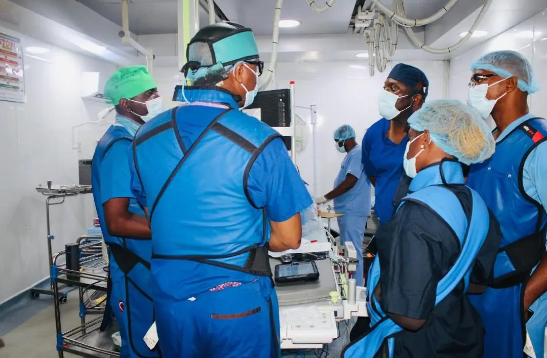
Coronary angiography: what to expect
An angiogram shows the cardiology team where heart arteries are narrowed or blocked.
Understanding coronary angiography
Coronary angiography is a procedure that lets the cardiology team see exactly where the arteries supplying your heart are narrowed or blocked, helping to decide between medicines, angioplasty, surgery or continued monitoring.
The heart is a muscle, and like any muscle it needs its own blood supply — delivered through the coronary arteries, which sit on the surface of the heart. Over time, fatty deposits can build up inside these arteries, a process called atherosclerosis, narrowing the space blood flows through. Symptoms such as chest pain, breathlessness or an abnormal stress test can point to this happening, but they cannot show precisely where or how severe a narrowing is. Angiography answers that question directly: a thin, flexible catheter is guided through a blood vessel, usually entering at the wrist or groin, and threaded to the opening of the coronary arteries. A contrast dye is then injected and tracked with continuous X-ray imaging, outlining the inside of the arteries and revealing any narrowing or blockage in real time.
When this procedure is recommended
- ●Often recommended after concerning symptoms such as chest pain, or after an abnormal stress test, ECG or other heart investigation
- ●May be planned in advance, or performed urgently when a heart attack is suspected
- ●Used to guide the choice between medicines, angioplasty and stenting, bypass surgery, or continued monitoring
- ●Sometimes used to reassess arteries after previous treatment, or before planned heart surgery for another reason
How to prepare
- ✓Follow fasting and medicine-pause instructions exactly — some blood thinners and diabetes medicines may need adjusting beforehand
- ✓Tell the team about kidney problems, or any previous reaction to contrast dye or anaesthesia
- ✓Share a full list of medicines, including herbal and over-the-counter products
- ✓Arrange transport and support as advised — you should not drive yourself home
- ✓Plan for the possibility of an overnight stay, even where a same-day discharge is expected
What happens on the day
You are usually admitted a few hours beforehand for final checks, consent and preparation of the entry site, most often at the wrist. In the procedure room, you lie on a table under continuous heart-rhythm and blood-pressure monitoring throughout.
A local anaesthetic numbs the entry site before the catheter is introduced — you should feel pressure rather than sharp pain. Most patients have light sedation to help them relax but remain awake and able to talk to the team. You may notice a brief warm flushing feeling when the contrast dye is injected; this is normal and passes within moments. The procedure itself is usually completed within around thirty minutes to an hour, though this varies depending on what is found and whether treatment is needed at the same time.
Afterward, firm pressure is applied to the entry site, sometimes with a small closure device at the groin, and you move to a recovery area where your heart rhythm, blood pressure and access site are checked regularly before you are cleared to go home or to a ward.
How the images become a plan
The cardiologist reviews the moving X-ray images during the procedure and can often tell you the key findings before you leave the room, though the full clinical picture — combined with your symptoms, other test results and overall health — is usually discussed at a follow-up review.
Depending on what is found, three broad paths are possible: no significant blockage, meaning symptoms likely have another explanation; a narrowing suitable for treatment with a balloon and stent in the same or a later procedure; or disease that is better suited to bypass surgery or continued medical treatment. Your clinician will explain which applies to you and why.
How Cardiocare prepares and assesses you
The exact pathway depends on your symptoms, examination, previous results and overall health. It typically includes:
- 01Pre-procedure review, consent and blood tests. Including kidney function and blood count before contrast dye is used.
- 02Medication and allergy check. Particularly blood thinners, diabetes medicines, and any previous contrast reaction.
- 03Catheter-based imaging in the cath lab. The angiography itself, under continuous monitoring.
- 04Observation of the access site and vital signs afterward. Before you are cleared for discharge or transfer to a ward.
Is this an emergency?
Right now
- After discharge: heavy bleeding, a rapidly swelling access site, severe chest pain, fainting or sudden breathlessness
- A cold, pale or painful limb below the access site
- Fever with chills
Go to the nearest emergency department immediately — do not drive yourself. Call Cardiocare's 24/7 emergency line on +234 806 142 4614 (staffed around the clock) to tell us you are coming.
Call Cardiocare 24/7Book sooner
Contact the team promptly for gradually increasing bruising, mild persistent oozing at the access site, or new symptoms that concern you but do not match the emergency list above.
Routine review
Mild bruising and tenderness at the access site that is settling as expected can usually be discussed at your scheduled follow-up.
Preparing for your appointment
What to bring
- ✓A list of all medicines you take, including herbal and over-the-counter preparations
- ✓Any previous ECGs, stress test results or hospital letters
- ✓Your HMO card and a valid means of identification
- ✓Contact details for the person bringing you home
What we will ask
Your symptoms and how they affect you, your full medicine and allergy history, any kidney problems, and any previous reaction to contrast dye or anaesthesia.
Questions worth asking us
- ●What are you hoping to find, and what happens if you find it?
- ●Could treatment happen in the same procedure if a blockage is found?
- ●What are the alternatives, including not having the procedure?
- ●How long will I need to stay, and what will recovery involve?